Our heart is a muscle that pumps blood through our veins, carrying the necessary oxygen to all parts of our body. Without the regular blood flow circulation that is enabled by the heart, which serves as a pump, the body can’t survive.
In cardiac arrest cases, CPR is recommended in the very first minutes because, after 8 minutes, the possibility of severe organ and brain damage and death is extremely high.
That’s why it’s important to understand what happens inside our body and how does CPR work physiologically. In this article, we will take a closer look into the cardiovascular system and explain how it works as well as what happens during CPR.
The Human Cardiovascular System
Our cardiovascular system is a complex and punctual mechanism of interconnected cells and organs that is responsible for transporting oxygen-rich blood into the body cells and transporting the CO2 blood back to the heart. It’s called cardio-vascular, from the ancient Greek terms “kardia” meaning “heart”, and the Latin “vascularis”, standing for “vessels” or “tubes”.
The Structure of The Cardiovascular System
The entire cardiovascular system consists of many bigger and smaller parts, functioning in synchronization to circulate blood throughout the body with the help of the heart. Aside from the main organ, the heart, a closed web used as a transportation system for carrying blood in and out, is spread throughout our body. Depending on their size and function, these vessels are:
- Arteries– the blood vessels that carry out the oxygenated blood
- Veins – the vessels that take back the CO2 blood to the heart
- Capillaries – the tiniest blood vessels that branch out to the furthest parts of the body and the smallest cells
Moreover, it’s of crucial meaning to understand that the lungs and their alveoli should never fill with blood, like the rest of our cells. Thus, we further distinguish the regular blood circulation system – the systemic and the pulmonary.
The second one is a smaller system because it’s only used for exchanging oxygen and carbon dioxide between the vessels and the lungs, while the first transports the nutrient-rich blood to the body cells and back.
Structure of The Heart
The heart is a muscle that consists of 2 upper and 2 lower chambers – the atria and the ventricles. The lower ones are filled with CO2 blood, while the upper two with oxygenated blood.
These must never meet and are naturally separated by a septum wall made out of elastic muscle tissue. The elastic valves open up and close down at a regulated tempo to keep up the regular cycle.
How Does Blood Flow Through Our Bodies?
Now that we’ve cleared out the pathways and organs responsible for pumping the blood, it’s time to understand the actual process of how it happens.
This is called “the cardiac cycle”, and it consists of two separate phases:
- The diastole, which is when the blood vessels return the blood to the heart, and the next contraction rapidly start the second phase.
- The systole where the “fresh” blood ejects out of the heart under increased pressure.
In reality, these phases can’t really be distinguished from one another, and this is how the entire blood flow goes.
- The CO2 blood returns back to the right upper heart chamber through the main vein – the vena cava.
- It then flows through, entering the lower right chamber, from where it’s pumped out of the heart through the pulmonary artery.
- The CO2 blood enters the lungs (the small capillaries circling the alveoli), where it’s replaced with oxygen-rich blood. This is done thanks to the natural difference in the molecular and mass weight of O2 and CO2.
- The oxygen-rich blood enters the heart through the upper left chamber through 4 pulmonary vessels.
- Entering the lower left chamber, the oxygenated blood leaves the heart through the main aorta.
As you may conclude, the heart’s cycle is reverse-clockwise, but in reality, it’s more punctual than any human-made mechanism.
The Importance of CPR for A Proper Heart Cycle
CPR stands for cardiopulmonary resuscitation, and it’s one of the most common life-saving techniques in cases of sudden cardiac or respiratory arrest. It’s essential to start performing CPR within mere minutes to increase the chances of survival of the unconscious person.
Until the 911 gets there, which is 7-14 minutes on average, depending on the region, using hand pressure and compressions on the person’s chest are very important. This is either to clear the airway and force the excess water and liquids if the victim is drowning and allow regular ventilation, or, in case of cardiac arrest, to try squeezing the blood left in the heart to start moving again.
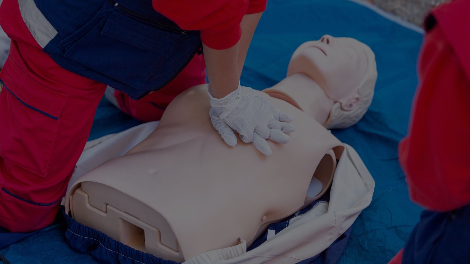
Manual CPR
CPR consists of a few basic movements of applying pressure to the person’s chest that is supposed to mimic the heartbeat and, in some cases, rescue breaths. It’s important to note that applying rapid and sharp pressure is more important than heavy pressure.
During CPR and closed chest compressions, compressions and decompressions are crucial to keep oxygen ventilation. Providing CPR within the first 1-4 minutes is very important during a cardiac arrest since it increases the chances of further defibrillation and successful resuscitation.
But what actually happens when we are compressing the person’s chest? CPR’s physiology can be understood through two basic concepts:
The Cardiac Pump Model
The direct compression of the heart leads to blood getting squeezed out of the heart, making way for fresh, oxygenated blood to enter. Since the main cause of cardiac arrest is the termination of the heart’s normal movements and compressions, squeezing the middle of the heart bone can serve as a pump to activate this process by using external force.
The Thoracic Pump Model
The thoracic model is the negative chest pressure, where the body releases oxygen. In the case of CPR, it would be the decompression phase, which is crucial to keeping the blood flow and airflow in the lungs. This causes fresh oxygenated blood to circulate persistently because the decompressions are necessary to pull out the carbonized blood into the vena cava, which is located on the right side of the heart, and find its way back to the lungs, where it will be exchanged with fresh, oxygenated blood.
CPR using an AED
If the event is in a crowded, public place, you should search for an AED – Automatic Electric Defibrillator. Like the electrocardiogram (ECG), the AED measures the heart’s rhythm, however, when necessary, sends a shock to the heart (also called defibrillation) to help the heart get back to a normal rhythm. The combined use of both CPR and AED is recommended to increase the chances of survival.
The Decompression Phase of CPR
The heart presses and decompresses in its natural way of working. Thus, paying attention to applying pressure is just as important as letting the chest and internal organs return to their normal position.
If you don’t pay attention to equalizing both cycles, chances are that internal organs might suffer from internal bleeding and additionally worsen what already appears as a bad situation.
Wrapping Up
Keeping up with the tempo and not forgetting about the phase of decompression is what a person performing CPR needs to remember. By regularly applying pressure within a timely tempered interval, CPR mimics normal heart movement.
As such, some of the internal chambers and valves might be forced to open, or some additional blood that stands inside the blood vessels will move and force further movement in the complex cardiovascular system.
This is the most assumed scientific explanation of what happens inside our body and how does CPR work physiologically. Whatever the case may be, CPR increases the chances of survival, especially in patients that experience these problems outside of the hospital. It’s important for cardiac arrest victims to receive high-quality CPR until the healthcare professionals arrive and take over to avoid brain damage or potential death.